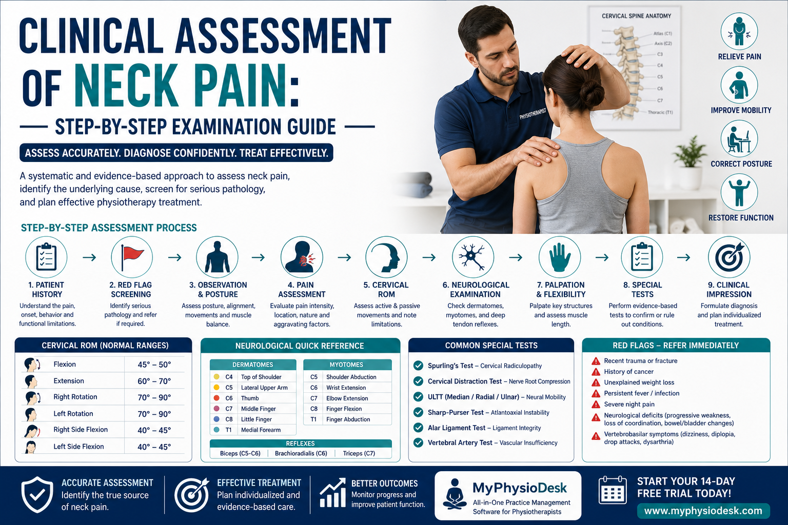
Clinical Assessment of Neck Pain: A Step-by-Step Examination Guide for Physiotherapists
Introduction
Neck pain is one of the most common musculoskeletal disorders encountered in physiotherapy practice and is a leading cause of disability worldwide. It affects people of all age groups, from office workers and students to athletes and elderly individuals. With increasing smartphone use, prolonged computer work, poor posture, and sedentary lifestyles, the prevalence of mechanical neck pain continues to rise.
Although most cases of neck pain are mechanical in origin, physiotherapists must always perform a systematic clinical assessment to identify the underlying cause, recognize serious pathology, determine contributing factors, and develop an individualized rehabilitation program.
A structured assessment improves diagnostic accuracy, guides evidence-based treatment, monitors patient progress, and enhances clinical decision-making throughout rehabilitation.
This comprehensive guide explains the essential steps every physiotherapist should follow while assessing a patient presenting with neck pain.
Understanding Cervical Spine Anatomy
Before assessing a patient, it is important to understand the anatomy and biomechanics of the cervical spine.
The cervical spine consists of seven vertebrae (C1–C7), intervertebral discs, facet joints, ligaments, muscles, and neural structures that work together to provide mobility, stability, and protection to the spinal cord.
The cervical spine is responsible for:
- Supporting the weight of the head.
- Allowing multidirectional movement.
- Protecting the spinal cord.
- Facilitating upper limb nerve function.
- Maintaining postural alignment.
Any dysfunction affecting these structures may result in pain, stiffness, muscle spasm, headaches, dizziness, or neurological symptoms.
Common Causes of Neck Pain
Neck pain may originate from various musculoskeletal, neurological, or systemic conditions. Understanding the possible causes helps narrow the differential diagnosis.
- Mechanical Neck Pain
- Poor Posture (Forward Head Posture)
- Cervical Muscle Strain
- Cervical Spondylosis
- Cervical Disc Herniation
- Cervical Radiculopathy
- Facet Joint Dysfunction
- Whiplash Injury
- Myofascial Pain Syndrome
- Upper Crossed Syndrome
- Cervicogenic Headache
- Thoracic Outlet Syndrome
Less commonly, neck pain may indicate serious pathology such as fractures, infections, inflammatory diseases, spinal cord compression, tumors, or vascular disorders. Therefore, every assessment should begin with appropriate screening.
Why a Structured Assessment is Important
A comprehensive assessment enables physiotherapists to identify the source of symptoms and distinguish mechanical neck pain from serious medical conditions.
A proper assessment helps to:
- Identify pain-generating structures.
- Differentiate musculoskeletal and neurological conditions.
- Screen for cervical radiculopathy and myelopathy.
- Recognize red flags requiring medical referral.
- Develop individualized treatment goals.
- Monitor patient progress objectively.
- Improve communication with other healthcare professionals.
Step 1: Patient History
A detailed subjective assessment often provides the most valuable diagnostic information. Before beginning the physical examination, gather a complete history of the patient's condition.
Chief Complaint
Ask the patient:
- What brings you to the clinic today?
- When did the pain begin?
- Was the onset sudden or gradual?
- Did any injury or accident occur?
History of Present Illness
- Duration of symptoms
- Pain progression
- Previous episodes
- Previous treatments
- Medication history
Pain Characteristics
- Location
- Radiating pain
- Severity
- Nature of pain (Sharp, Dull, Burning, Tingling)
- Morning stiffness
- Night pain
Aggravating Factors
- Looking down
- Computer work
- Driving
- Sleeping position
- Overhead activities
- Prolonged sitting
Relieving Factors
- Rest
- Heat therapy
- Medication
- Postural correction
- Movement
Step 2: Medical History
Review the patient's overall medical condition before proceeding with physical examination.
Important areas include:
- Previous cervical injuries
- Previous surgeries
- Osteoporosis
- Rheumatoid Arthritis
- Diabetes Mellitus
- Cancer History
- Recent infections
- Long-term steroid use
- Hypertension
- Neurological disorders
Step 3: Screen for Red Flags
Red flag screening is essential to identify patients who require urgent medical referral rather than physiotherapy management.
Immediately investigate if the patient reports:
- Recent major trauma
- Severe unrelenting night pain
- Unexplained weight loss
- History of cancer
- Persistent fever or infection
- Difficulty swallowing
- Loss of coordination
- Bilateral neurological symptoms
- Bowel or bladder dysfunction
- Progressive upper limb weakness
- Sudden dizziness or visual disturbances
If any red flags are identified, postpone physiotherapy treatment and refer the patient to the appropriate medical specialist immediately.
Step 4: Pain Assessment
Pain assessment should evaluate both the intensity and behavior of symptoms.
Use standardized outcome measures such as:
- Visual Analogue Scale (VAS)
- Numeric Pain Rating Scale (NPRS)
- Neck Disability Index (NDI)
Document:
- Pain intensity
- Pain location
- Radiation
- Frequency
- Irritability
- 24-hour pain pattern
- Functional limitations
Recording baseline pain scores helps measure improvement during future reassessment.
Step 5: Observation and Postural Assessment
Observation begins as soon as the patient enters the clinic. A skilled physiotherapist can identify numerous abnormalities before touching the patient.
Observe the patient while:
- Walking
- Sitting
- Standing
- Turning
- Removing clothing
Postural Assessment
Assess for:
- Forward Head Posture
- Rounded Shoulders
- Thoracic Kyphosis
- Scapular Position
- Shoulder Height Asymmetry
- Muscle Bulk
- Protective Guarding
- Head Tilt
- Cervical Alignment
Poor posture is one of the most common contributors to chronic mechanical neck pain. Document all postural abnormalities, as they often influence treatment planning and exercise prescription.
Clinical Tip
Never rely solely on imaging findings. Many patients with cervical degeneration or disc bulges on MRI may have minimal symptoms, while others with normal imaging may experience significant pain. Always correlate imaging results with your clinical examination and functional assessment before reaching a diagnosis.
Step 6: Cervical Range of Motion (ROM) Assessment
After completing the subjective examination and postural assessment, evaluate the patient's cervical range of motion (ROM). ROM assessment helps identify movement restrictions, painful directions, muscle tightness, joint dysfunction, and potential nerve involvement.
Assess Active Cervical Movements first:
- Flexion
- Extension
- Right Rotation
- Left Rotation
- Right Side Flexion
- Left Side Flexion
Observe:
- Range of movement
- Pain during movement
- Movement quality
- Compensatory trunk movement
- Muscle guarding
- End feel
If active movement is limited, carefully assess passive physiological movements to determine whether the limitation is muscular, capsular, or joint-related.
Normal Cervical Range of Motion
| Movement | Normal Range |
|---|---|
| Flexion | 45–50° |
| Extension | 60–70° |
| Rotation | 70–90° |
| Side Flexion | 40–45° |
Always compare movement on both sides and note any asymmetry.
Step 7: Neurological Examination
Patients presenting with radiating neck pain, numbness, tingling, muscle weakness, or altered sensation require a comprehensive neurological examination.
The neurological examination helps determine whether cervical nerve root compression, spinal cord involvement, or peripheral nerve pathology is present.
Dermatomes Assessment
Assess light touch sensation over the corresponding cervical dermatomes.
| Nerve Root | Area Tested |
|---|---|
| C4 | Top of Shoulder |
| C5 | Lateral Upper Arm |
| C6 | Thumb |
| C7 | Middle Finger |
| C8 | Little Finger |
| T1 | Medial Forearm |
Compare both sides for altered sensation or numbness.
Myotome Assessment
Manual Muscle Testing (MMT) helps identify weakness caused by cervical nerve root compression.
| Nerve Root | Muscle Action |
|---|---|
| C5 | Shoulder Abduction |
| C6 | Elbow Flexion / Wrist Extension |
| C7 | Elbow Extension |
| C8 | Finger Flexion |
| T1 | Finger Abduction |
Deep Tendon Reflexes
Assess reflexes using a reflex hammer.
| Reflex | Nerve Root |
|---|---|
| Biceps | C5–C6 |
| Brachioradialis | C6 |
| Triceps | C7 |
Hyperreflexia may indicate cervical myelopathy, whereas diminished reflexes may suggest cervical radiculopathy.
Step 8: Palpation
Palpation should always be gentle and systematic.
Assess the following structures:
- Spinous Processes
- Facet Joints
- Upper Trapezius
- Levator Scapulae
- Sternocleidomastoid (SCM)
- Scalenes
- Suboccipital Muscles
- Paraspinal Muscles
- Supraspinous Ligament
Observe for:
- Tenderness
- Muscle Spasm
- Trigger Points
- Swelling
- Temperature Changes
- Muscle Tightness
Step 9: Muscle Length Assessment
Shortened muscles frequently contribute to chronic neck pain and postural dysfunction.
Assess flexibility of:
- Upper Trapezius
- Levator Scapulae
- Pectoralis Major
- Pectoralis Minor
- Sternocleidomastoid
- Scalenes
- Suboccipital Muscles
Muscle shortening often accompanies Forward Head Posture and Upper Crossed Syndrome.
Step 10: Functional Assessment
The patient's functional limitations often provide more useful information than pain alone.
Ask the patient about difficulty with:
- Driving
- Computer Work
- Mobile Phone Use
- Reading
- Sleeping
- Looking Up
- Lifting Objects
- Overhead Activities
- Household Work
- Sports Activities
Functional assessment helps prioritize treatment goals based on activities that matter most to the patient.
Outcome Measures
Use validated outcome measures to objectively evaluate disability and monitor progress.
Recommended questionnaires include:
- Neck Disability Index (NDI)
- Patient-Specific Functional Scale (PSFS)
- Visual Analogue Scale (VAS)
- Numeric Pain Rating Scale (NPRS)
Repeat these assessments during follow-up visits to objectively monitor improvement.
Clinical Pearl
Always correlate neurological findings with the patient's symptoms. A reduced reflex or mild sensory deficit without matching clinical symptoms may not always indicate cervical nerve root compression. Clinical reasoning should combine subjective findings, physical examination, neurological testing, and functional assessment before reaching a diagnosis.
Step 11: Special Tests
Special tests should be performed only after completing the subjective assessment, neurological examination, and cervical range of motion assessment. These tests help confirm or rule out specific cervical pathologies and should always be interpreted alongside the patient's history and clinical findings.
1. Spurling's Test
Purpose: Detect cervical nerve root compression (Cervical Radiculopathy).
Procedure:
- Patient sits comfortably.
- Extend the cervical spine.
- Side bend and rotate the head toward the symptomatic side.
- Apply gentle downward compression.
Positive Test:
- Radiating pain into the arm.
- Paresthesia following a dermatome.
2. Cervical Distraction Test
Purpose: Identify cervical nerve root compression.
Procedure:
- Patient lies supine.
- Therapist gently distracts the cervical spine.
Positive Test:
- Reduction of neck or arm pain.
3. Upper Limb Tension Test (ULTT)
Purpose: Assess neural tissue mobility and nerve irritation.
The test helps identify:
- Median Nerve
- Radial Nerve
- Ulnar Nerve
Positive Test:
- Reproduction of familiar symptoms.
- Restricted neural mobility.
4. Sharp-Purser Test
Purpose: Evaluate atlantoaxial instability.
Positive Test:
- Reduction of neurological symptoms.
- Excessive cervical movement.
This test should only be performed by trained clinicians when instability is suspected.
5. Alar Ligament Stress Test
Purpose: Assess integrity of the alar ligaments.
Positive Test:
- Excessive movement.
- Delayed movement of C2.
6. Vertebral Artery Test
Purpose: Screen for vertebrobasilar insufficiency before cervical mobilization or manipulation.
Watch carefully for:
- Dizziness
- Diplopia
- Dysarthria
- Dysphagia
- Drop attacks
- Nausea
- Nystagmus
Step 12: Differential Diagnosis
After completing the examination, identify the most likely clinical diagnosis.
Common differential diagnoses include:
- Mechanical Neck Pain
- Cervical Radiculopathy
- Cervical Myelopathy
- Cervical Disc Herniation
- Facet Joint Dysfunction
- Whiplash Associated Disorder
- Upper Crossed Syndrome
- Cervicogenic Headache
- Thoracic Outlet Syndrome
- Myofascial Pain Syndrome
Step 13: Clinical Impression
Combine subjective findings, physical examination, neurological assessment, posture analysis, special tests, and functional limitations to establish a working physiotherapy diagnosis.
Remember that imaging findings should support—not replace—clinical reasoning.
Step 14: Develop an Individualized Treatment Plan
Based on the assessment findings, establish patient-centered rehabilitation goals.
Your treatment plan may include:
- Patient Education
- Postural Correction
- Ergonomic Advice
- Manual Therapy
- Cervical Mobilization (when appropriate)
- Stretching Exercises
- Deep Cervical Flexor Training
- Scapular Stabilization Exercises
- Strengthening Program
- Neural Mobilization
- Home Exercise Program
- Pain Management Strategies
Reassessment
Clinical reassessment is essential to monitor treatment effectiveness and modify rehabilitation plans.
Reassess:
- Pain Intensity
- Cervical ROM
- Muscle Strength
- Neurological Findings
- Functional Ability
- Neck Disability Index
- Patient Goals
Common Assessment Mistakes
- Ignoring patient history.
- Skipping red flag screening.
- Failing to assess posture.
- Not performing neurological examination.
- Over-relying on MRI findings.
- Ignoring functional limitations.
- Incomplete documentation.
- Not reassessing patient progress.
- Using painful techniques unnecessarily.
- Failing to educate the patient.
Clinical Assessment Checklist
- ✅ Patient History
- ✅ Pain Assessment
- ✅ Medical History
- ✅ Red Flag Screening
- ✅ Observation
- ✅ Postural Assessment
- ✅ Cervical ROM
- ✅ Neurological Examination
- ✅ Dermatomes
- ✅ Myotomes
- ✅ Reflexes
- ✅ Palpation
- ✅ Muscle Length Testing
- ✅ Functional Assessment
- ✅ Special Tests
- ✅ Clinical Diagnosis
- ✅ Treatment Goals
- ✅ Home Exercise Program
- ✅ Documentation
- ✅ Follow-up Plan
Evidence-Based Practice
Research supports the use of standardized assessment procedures and validated outcome measures when evaluating patients with neck pain. Combining clinical reasoning with objective findings improves diagnostic accuracy, treatment effectiveness, and patient satisfaction.
A thorough assessment also enables physiotherapists to detect serious pathology early, select appropriate interventions, and monitor rehabilitation outcomes more effectively.
Final Thoughts
Clinical assessment is the cornerstone of successful neck pain management. A systematic approach that includes patient history, red flag screening, postural assessment, cervical range of motion, neurological examination, special tests, and functional evaluation enables physiotherapists to make informed clinical decisions and deliver evidence-based care.
Consistent documentation is equally important. Accurate assessment records improve communication, support clinical reasoning, and ensure continuity of care throughout the rehabilitation process.
MyPhysioDesk helps physiotherapists simplify this process by providing built-in Digital Assessment Forms, structured SOAP Notes, Patient Progress Tracking, and secure Cloud-Based Patient Records. These tools make it easy to document every examination, compare findings across follow-up visits, and spend more time delivering quality patient care instead of managing paperwork.
Enhance Your Clinical Documentation with MyPhysioDesk
Whether you're treating mechanical neck pain, cervical radiculopathy, postural syndrome, or cervical disc disorders, MyPhysioDesk helps you organize assessments, maintain professional SOAP Notes, record outcome measures, monitor patient recovery, and manage your physiotherapy practice efficiently from one secure cloud-based platform.
Start your 14-Day FREE Trial today.
🌐 https://myphysiodesk.com
Back to blog