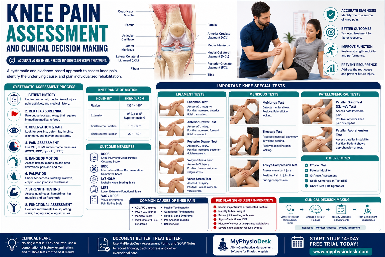
Knee Pain Assessment and Clinical Decision Making: A Complete Guide for Physiotherapists
Introduction
Knee pain is one of the most frequently encountered musculoskeletal complaints in physiotherapy practice, affecting individuals of all ages—from young athletes with ligament injuries to older adults with degenerative joint conditions. Because the knee is a complex weight-bearing joint, pain may originate from bones, ligaments, menisci, cartilage, tendons, muscles, bursae, or even referred pain from the hip or lumbar spine.
An accurate diagnosis cannot be made by imaging alone. A systematic clinical assessment enables physiotherapists to identify the source of symptoms, recognize serious pathology, prioritize functional limitations, and develop individualized rehabilitation programs based on evidence-based clinical reasoning.
This comprehensive guide outlines the step-by-step process of assessing patients with knee pain and provides practical insights that every physiotherapist can apply in daily clinical practice.
Understanding Knee Anatomy
The knee is the largest synovial joint in the human body and plays a vital role in walking, running, climbing stairs, squatting, jumping, and maintaining balance. Although designed primarily as a hinge joint, it also allows slight rotational movements essential for normal gait.
The knee joint consists of:
- Femur
- Tibia
- Patella
- Fibula (supporting structure)
Important stabilizing structures include:
- Anterior Cruciate Ligament (ACL)
- Posterior Cruciate Ligament (PCL)
- Medial Collateral Ligament (MCL)
- Lateral Collateral Ligament (LCL)
- Medial Meniscus
- Lateral Meniscus
- Patellar Tendon
- Quadriceps Tendon
- Joint Capsule
- Articular Cartilage
Understanding these anatomical structures helps physiotherapists correlate symptoms with potential pathology during assessment.
Common Causes of Knee Pain
Knee pain may result from traumatic injuries, overuse syndromes, degenerative conditions, inflammatory diseases, or biomechanical abnormalities.
Common clinical conditions include:
- Anterior Cruciate Ligament (ACL) Injury
- Posterior Cruciate Ligament (PCL) Injury
- Medial Collateral Ligament (MCL) Injury
- Lateral Collateral Ligament (LCL) Injury
- Meniscal Tear
- Patellofemoral Pain Syndrome
- Patellar Tendinopathy (Jumper's Knee)
- Quadriceps Tendinopathy
- Iliotibial Band Syndrome
- Pes Anserine Bursitis
- Osteoarthritis
- Rheumatoid Arthritis
- Osgood-Schlatter Disease
- Baker's Cyst
- Referred Pain from Hip or Lumbar Spine
Why Clinical Assessment is Important
A comprehensive knee assessment enables physiotherapists to differentiate between ligament injuries, meniscal pathology, patellofemoral disorders, tendon injuries, and degenerative conditions. It also helps identify patients who require urgent orthopedic referral.
A structured assessment helps to:
- Identify pain-generating structures.
- Recognize instability.
- Detect meniscal injuries.
- Assess functional limitations.
- Screen for serious pathology.
- Guide treatment planning.
- Monitor rehabilitation progress objectively.
Step 1: Patient History
A detailed subjective assessment often provides the most valuable diagnostic information before the physical examination begins.
Chief Complaint
- When did your knee pain begin?
- Which knee is affected?
- Was there any injury or trauma?
- Did symptoms develop suddenly or gradually?
Pain Characteristics
- Location of pain
- Pain intensity (VAS/NPRS)
- Sharp, dull, aching, burning, or stabbing pain
- Clicking or locking sensation
- Swelling
- Morning stiffness
- Night pain
Mechanism of Injury
Ask whether symptoms occurred during:
- Twisting movement
- Pivoting
- Landing after a jump
- Direct impact
- Running
- Climbing stairs
- Squatting
- Prolonged sitting
Aggravating Factors
- Walking
- Running
- Stair climbing
- Squatting
- Kneeling
- Jumping
- Sports participation
Relieving Factors
- Rest
- Ice application
- Medication
- Knee brace
- Activity modification
Previous Medical History
- Previous knee injuries
- Previous surgeries
- Sports participation
- Osteoarthritis
- Inflammatory arthritis
- Diabetes Mellitus
- Previous physiotherapy
Step 2: Red Flag Screening
Before beginning physical examination, screen for conditions requiring urgent medical referral.
Red flags include:
- Recent major trauma
- Suspected fracture
- Severe joint deformity
- Inability to bear weight
- Persistent fever
- Hot, swollen joint
- History of cancer
- Unexplained weight loss
- Severe night pain
- Signs of infection
- Deep Vein Thrombosis (DVT) symptoms
Patients presenting with suspected fractures, septic arthritis, acute joint dislocation, neurovascular compromise, or symptoms suggestive of deep vein thrombosis should be referred immediately for emergency medical evaluation before physiotherapy treatment is initiated.
Step 3: Observation
Observation begins the moment the patient enters the clinic. Careful visual assessment provides valuable information regarding gait, posture, swelling, and functional movement.
Observe during:
- Walking
- Standing
- Sitting
- Transfers
- Stair negotiation (if appropriate)
Look for:
- Antalgic gait
- Limping
- Knee valgus
- Knee varus
- Genu recurvatum
- Joint swelling
- Muscle wasting
- Bruising
- Scars from previous surgery
- Patellar alignment
Step 4: Pain Assessment
Pain assessment should evaluate both symptom severity and functional impact.
Recommended outcome measures include:
- Visual Analogue Scale (VAS)
- Numeric Pain Rating Scale (NPRS)
- Knee Injury and Osteoarthritis Outcome Score (KOOS)
- International Knee Documentation Committee (IKDC) Score
- Lysholm Knee Scoring Scale
Document:
- Pain intensity
- Pain location
- Swelling
- Pain during movement
- Morning stiffness
- Night pain
- Functional limitations
Step 5: Range of Motion (ROM) Assessment
Assess both active and passive knee movements to identify restrictions caused by pain, swelling, muscle tightness, or joint pathology.
Assess:
- Knee Flexion
- Knee Extension
- Tibial Rotation (when indicated)
Observe:
- Pain during movement
- Movement quality
- End feel
- Extension lag
- Flexion restriction
- Crepitus
- Joint locking
Normal Knee Range of Motion
| Movement | Normal ROM |
|---|---|
| Flexion | 135–145° |
| Extension | 0° (up to 5° hyperextension may be normal) |
Always compare the affected knee with the unaffected side and document any differences in movement, pain, or end feel.
Clinical Tip
A patient reporting a twisting injury followed by immediate swelling, instability, and a "popping" sensation should raise suspicion of an ACL injury. Conversely, locking, clicking, and joint line pain after a twisting movement often suggest a meniscal tear. Clinical reasoning should always integrate the patient's history with objective examination findings.
Step 6: Palpation
Palpation is performed after observation and range of motion assessment to identify tenderness, swelling, joint effusion, warmth, crepitus, and abnormalities of the soft tissues surrounding the knee. Compare findings with the unaffected side to improve diagnostic accuracy.
Systematically palpate the following structures:
- Patella
- Quadriceps Tendon
- Patellar Tendon
- Tibial Tuberosity
- Medial Joint Line
- Lateral Joint Line
- Medial Collateral Ligament (MCL)
- Lateral Collateral Ligament (LCL)
- Fibular Head
- Pes Anserinus
- Iliotibial Band
- Popliteal Fossa
- Baker's Cyst Region
Observe for:
- Localized tenderness
- Joint line pain
- Effusion
- Warmth
- Soft tissue swelling
- Muscle wasting
- Crepitus
Step 7: Muscle Strength Assessment
Manual Muscle Testing (MMT) helps identify weakness caused by ligament injuries, tendon disorders, pain inhibition, neurological conditions, or prolonged immobilization.
| Movement | Primary Muscle | Nerve Supply |
|---|---|---|
| Knee Extension | Quadriceps | Femoral Nerve |
| Knee Flexion | Hamstrings | Sciatic Nerve |
| Hip Abduction | Gluteus Medius | Superior Gluteal Nerve |
| Hip Extension | Gluteus Maximus | Inferior Gluteal Nerve |
| Calf Raise | Gastrocnemius & Soleus | Tibial Nerve |
Weakness of the quadriceps and hip abductors is frequently associated with patellofemoral pain syndrome and chronic knee dysfunction.
Step 8: Functional Assessment
Evaluate how knee pain affects the patient's daily activities. Functional limitations often provide more clinically relevant information than pain intensity alone.
Assess the patient's ability to perform:
- Walking
- Running
- Squatting
- Lunging
- Sit-to-Stand
- Single Leg Standing
- Stair Climbing
- Stair Descent
- Jumping
- Landing
Observe movement quality, dynamic knee alignment, pain reproduction, instability, and compensatory movement patterns.
Ligament Stability Tests
Ligament testing evaluates the integrity of the major stabilizing structures of the knee. These tests should be interpreted together with the patient's injury mechanism and clinical history.
1. Lachman Test
Purpose: Assess Anterior Cruciate Ligament (ACL) integrity.
Procedure:
- Patient lies supine.
- Flex the knee approximately 20–30°.
- Stabilize the femur with one hand.
- Pull the tibia anteriorly with the other hand.
Positive Test:
- Excessive anterior tibial translation.
- Soft or absent end feel.
The Lachman Test is considered one of the most sensitive clinical tests for ACL injury.
2. Anterior Drawer Test
Purpose: Assess ACL injury.
Procedure:
- Flex the hip to 45°.
- Flex the knee to 90°.
- Stabilize the foot.
- Pull the tibia forward.
Positive Test:
- Increased anterior movement of the tibia.
3. Posterior Drawer Test
Purpose: Assess Posterior Cruciate Ligament (PCL).
Procedure:
- Position the knee at 90° flexion.
- Push the tibia posteriorly.
Positive Test:
- Excessive posterior tibial translation.
4. Valgus Stress Test
Purpose: Evaluate Medial Collateral Ligament (MCL).
Procedure:
- Flex the knee approximately 30°.
- Apply a valgus force.
Positive Test:
- Medial joint pain.
- Excessive joint opening.
5. Varus Stress Test
Purpose: Evaluate Lateral Collateral Ligament (LCL).
Procedure:
- Flex the knee approximately 30°.
- Apply a varus force.
Positive Test:
- Lateral joint pain.
- Excessive lateral joint opening.
Meniscal Assessment
Meniscal injuries commonly occur following twisting movements and frequently present with joint line pain, locking, clicking, or episodes of giving way.
6. McMurray Test
Purpose: Detect meniscal tears.
Procedure:
- Flex the knee completely.
- Rotate the tibia while extending the knee.
Positive Test:
- Pain.
- Palpable or audible click.
- Joint locking.
7. Thessaly Test
Purpose: Assess meniscal pathology during weight-bearing.
Procedure:
- Patient stands on one leg.
- Flex the knee approximately 20°.
- Rotate the body internally and externally.
Positive Test:
- Joint line pain.
- Locking sensation.
- Mechanical symptoms.
8. Apley's Compression Test
Purpose: Assess meniscal injury.
Procedure:
- Patient lies prone.
- Flex the knee to 90°.
- Apply downward compression while rotating the tibia.
Positive Test:
- Joint line pain during compression.
Patellofemoral Assessment
Patellofemoral disorders are a common cause of anterior knee pain, particularly in runners, athletes, and young adults.
9. Patellar Grind Test (Clarke's Sign)
Purpose: Assess patellofemoral joint dysfunction.
Procedure:
- Patient lies supine.
- Apply gentle downward pressure over the superior pole of the patella.
- Ask the patient to contract the quadriceps.
Positive Test:
- Anterior knee pain.
- Inability to maintain quadriceps contraction.
10. Patellar Apprehension Test
Purpose: Evaluate patellar instability.
Procedure:
- Patient lies supine with the knee slightly flexed.
- Gently translate the patella laterally.
Positive Test:
- Patient demonstrates apprehension.
- Guarding or fear of dislocation.
Clinical Pearl
No individual ligament or meniscal test is perfectly accurate. The most reliable diagnosis comes from combining patient history, injury mechanism, palpation findings, functional assessment, and clusters of special tests. Clinical reasoning is always more valuable than relying on a single positive test.
Step 11: Functional Outcome Measures
Standardized outcome measures provide objective data to evaluate disability, monitor rehabilitation progress, and support evidence-based clinical decision making. These tools should be recorded during the initial assessment and repeated at regular intervals throughout rehabilitation.
Commonly used outcome measures include:
- Knee Injury and Osteoarthritis Outcome Score (KOOS)
- International Knee Documentation Committee (IKDC) Subjective Knee Form
- Lysholm Knee Scoring Scale
- Tegner Activity Scale
- Lower Extremity Functional Scale (LEFS)
- Visual Analogue Scale (VAS)
- Numeric Pain Rating Scale (NPRS)
Using standardized questionnaires allows physiotherapists to objectively measure pain, symptoms, functional limitations, sports participation, and quality of life while demonstrating treatment effectiveness.
Step 12: Differential Diagnosis
After completing the subjective and objective examination, physiotherapists should integrate all findings to determine the most likely diagnosis. Knee pain may arise from multiple anatomical structures, making differential diagnosis an essential component of clinical reasoning.
Common differential diagnoses include:
- Anterior Cruciate Ligament (ACL) Tear
- Posterior Cruciate Ligament (PCL) Tear
- Medial Collateral Ligament (MCL) Injury
- Lateral Collateral Ligament (LCL) Injury
- Medial Meniscus Tear
- Lateral Meniscus Tear
- Patellofemoral Pain Syndrome
- Patellar Tendinopathy
- Quadriceps Tendinopathy
- Osteoarthritis of the Knee
- Pes Anserine Bursitis
- Iliotibial Band Syndrome
- Baker's Cyst
- Referred Pain from the Hip
- Lumbar Radiculopathy
Step 13: Clinical Decision Making
Clinical decision making is the process of combining patient history, physical examination findings, functional assessment, and special test results to identify the primary pain source and determine the most appropriate rehabilitation strategy.
Examples of evidence-based clinical reasoning include:
- Twisting injury + immediate swelling + positive Lachman Test → Suspect ACL Tear.
- Joint line tenderness + locking + positive McMurray Test → Suggest Meniscal Tear.
- Anterior knee pain + pain during stair climbing + positive Clarke's Test → Suggest Patellofemoral Pain Syndrome.
- Pain below the patella during jumping + tenderness over patellar tendon → Suggest Patellar Tendinopathy.
- Medial knee pain after valgus injury + positive Valgus Stress Test → Suggest MCL Injury.
Always correlate examination findings with the patient's symptoms and functional limitations rather than relying on a single positive test.
Step 14: Develop an Individualized Treatment Plan
Treatment should always be based on assessment findings, patient goals, and functional requirements.
Your rehabilitation program may include:
- Patient Education
- Pain Management Strategies
- Activity Modification
- Manual Therapy
- Joint Mobilization
- Quadriceps Strengthening
- Hamstring Strengthening
- Hip Strengthening Exercises
- Neuromuscular Control Training
- Balance and Proprioception Training
- Functional Movement Retraining
- Gait Training
- Sports-Specific Rehabilitation
- Home Exercise Program
Treatment intensity should progress according to pain levels, tissue healing, and functional improvement while ensuring patient safety.
Reassessment
Reassessment is essential throughout rehabilitation to evaluate treatment effectiveness and determine whether rehabilitation goals are being achieved.
Reassess:
- Pain Intensity (VAS/NPRS)
- Knee Range of Motion
- Muscle Strength
- Joint Swelling
- Ligament Stability
- Meniscal Signs
- Functional Performance
- KOOS
- IKDC Score
- Patient Goals
Objective reassessment enables physiotherapists to modify rehabilitation plans based on measurable patient progress.
Common Assessment Mistakes
- Skipping detailed patient history.
- Ignoring the mechanism of injury.
- Not screening for red flags.
- Performing only one special test.
- Ignoring hip and lumbar spine assessment.
- Over-relying on MRI findings.
- Failing to assess gait and functional movement.
- Incomplete documentation.
- Not reassessing progress objectively.
- Neglecting patient education and home exercise instruction.
Comprehensive Knee Assessment Checklist
- ✅ Patient History
- ✅ Pain Assessment
- ✅ Medical History
- ✅ Red Flag Screening
- ✅ Observation
- ✅ Gait Analysis
- ✅ Postural Assessment
- ✅ Knee ROM
- ✅ Palpation
- ✅ Muscle Strength Testing
- ✅ Functional Assessment
- ✅ Ligament Stability Tests
- ✅ Meniscal Tests
- ✅ Patellofemoral Assessment
- ✅ Outcome Measures
- ✅ Differential Diagnosis
- ✅ Clinical Impression
- ✅ Treatment Goals
- ✅ Home Exercise Program
- ✅ Documentation
- ✅ Follow-up Plan
Evidence-Based Practice
Current evidence recommends using clusters of clinical tests rather than relying on a single examination technique. Combining patient history, observation, ligament testing, meniscal assessment, functional performance, and validated outcome measures improves diagnostic accuracy and supports better rehabilitation outcomes.
Objective documentation also enhances communication with orthopedic surgeons, sports physicians, and other healthcare professionals involved in patient care.
Final Thoughts
A systematic knee assessment is the foundation of successful physiotherapy management. By integrating patient history, observation, palpation, range of motion assessment, muscle strength testing, functional evaluation, ligament stability tests, meniscal assessment, and evidence-based clinical reasoning, physiotherapists can accurately diagnose knee disorders and develop individualized rehabilitation programs that optimize patient outcomes.
Consistent documentation is equally important for monitoring recovery, comparing reassessment findings, and ensuring continuity of care throughout the rehabilitation process.
MyPhysioDesk simplifies knee assessment by providing built-in Digital Assessment Forms, structured SOAP Notes, Outcome Measure Recording, Goal Tracking, Patient Progress Monitoring, and secure Cloud-Based Patient Records. These tools help physiotherapists document every evaluation professionally, compare findings across follow-up visits, and focus more on delivering exceptional patient care instead of managing paperwork.
Deliver Better Knee Rehabilitation with MyPhysioDesk
Whether you're managing ligament injuries, meniscal tears, patellofemoral pain syndrome, osteoarthritis, sports injuries, or post-operative rehabilitation, MyPhysioDesk helps you organize assessments, create professional SOAP Notes, record outcome measures, monitor patient recovery, and manage your physiotherapy practice from one secure cloud-based platform.
Start your 14-Day FREE Trial today.
🌐 https://myphysiodesk.com
Back to blog