Shoulder Pain Assessment: Special Tests Every Physiotherapist Should Know
Introduction
Shoulder pain is one of the most common musculoskeletal complaints treated in physiotherapy practice. Due to the shoulder's remarkable mobility and complex anatomy, identifying the exact source of pain can often be challenging. Patients may present with pain during overhead activities, lifting, reaching behind the back, sports participation, or even simple daily activities such as dressing and grooming.
A successful rehabilitation program begins with a systematic and evidence-based clinical assessment. Rather than relying solely on imaging reports, physiotherapists should combine subjective examination, physical assessment, movement analysis, and special tests to determine the underlying pathology and develop an individualized treatment plan.
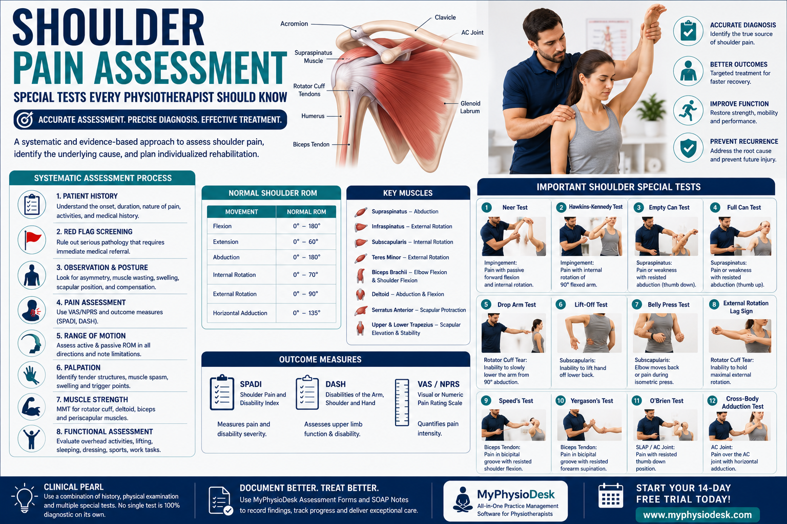
This comprehensive guide explains the essential assessment process and introduces the most reliable special tests every physiotherapist should know when evaluating patients with shoulder pain.
Understanding Shoulder Anatomy
The shoulder is the most mobile joint in the human body. It consists of multiple joints, muscles, ligaments, tendons, and bursae that work together to provide stability and movement.
The shoulder complex includes:
- Glenohumeral Joint
- Acromioclavicular (AC) Joint
- Sternoclavicular Joint
- Scapulothoracic Articulation
The primary stabilizing structures include:
- Rotator Cuff Muscles
- Glenoid Labrum
- Joint Capsule
- Glenohumeral Ligaments
- Long Head of Biceps Tendon
Because several anatomical structures can produce similar symptoms, a structured assessment is essential for accurate diagnosis.
Common Causes of Shoulder Pain
Shoulder pain may arise from muscles, tendons, joints, ligaments, nerves, or referred pain from the cervical spine.
Common conditions include:
- Rotator Cuff Tendinopathy
- Shoulder Impingement Syndrome
- Subacromial Bursitis
- Frozen Shoulder (Adhesive Capsulitis)
- Biceps Tendinitis
- SLAP Lesion
- Acromioclavicular Joint Injury
- Shoulder Instability
- Rotator Cuff Tear
- Calcific Tendinitis
- Glenohumeral Osteoarthritis
- Cervical Radiculopathy (Referred Pain)
Why a Structured Shoulder Assessment Matters
A detailed assessment enables physiotherapists to identify the pain-generating structure, determine functional limitations, recognize serious pathology, and select the most appropriate rehabilitation strategy.
A proper assessment helps to:
- Identify the primary source of pain.
- Differentiate between contractile and non-contractile tissues.
- Recognize cervical spine involvement.
- Identify instability and labral injuries.
- Detect rotator cuff pathology.
- Develop individualized treatment goals.
- Monitor rehabilitation progress objectively.
Step 1: Patient History
The subjective assessment often provides the most valuable information for diagnosing shoulder disorders. Allow the patient to describe symptoms in their own words before asking focused questions.
Chief Complaint
- When did your shoulder pain begin?
- Did the pain start suddenly or gradually?
- Was there any injury or trauma?
- Which shoulder is affected?
Pain Characteristics
- Pain location
- Pain intensity (VAS/NPRS)
- Sharp, dull, aching, burning, or throbbing pain
- Radiating symptoms
- Night pain
- Morning stiffness
Aggravating Activities
- Overhead reaching
- Lifting objects
- Throwing activities
- Dressing
- Sleeping on the affected shoulder
- Sports activities
Relieving Factors
- Rest
- Medication
- Ice or heat therapy
- Supporting the arm
- Avoiding overhead activities
Previous Medical History
- Previous shoulder injuries
- Previous surgeries
- Diabetes Mellitus
- Rheumatoid Arthritis
- Cervical Spine Disorders
- Previous Physiotherapy
Step 2: Red Flag Screening
Before beginning physical examination, physiotherapists must exclude serious pathology that requires urgent medical referral.
Watch for:
- Recent fracture or major trauma
- Unexplained weight loss
- History of cancer
- Persistent fever
- Severe night pain not relieved by rest
- Shoulder deformity
- Progressive neurological deficits
- Signs of infection
Patients presenting with significant trauma, suspected fracture, shoulder dislocation, infection, or unexplained neurological symptoms should be referred immediately for appropriate medical evaluation.
Step 3: Observation
Observation begins as soon as the patient enters the clinic. A careful visual examination often provides important diagnostic clues before physical testing begins.
Observe the patient during:
- Walking
- Sitting
- Standing
- Removing clothing
- Natural arm swing
Look for:
- Shoulder asymmetry
- Scapular winging
- Rounded shoulders
- Forward head posture
- Muscle wasting
- Swelling
- Bruising
- Deformity
- Protective positioning
Step 4: Postural Assessment
Postural abnormalities frequently contribute to chronic shoulder pain by altering scapular mechanics and increasing stress on the rotator cuff.
Assess:
- Head Position
- Cervical Alignment
- Thoracic Kyphosis
- Scapular Position
- Scapular Symmetry
- Shoulder Height
- Clavicle Position
Patients with Forward Head Posture and Rounded Shoulders commonly develop shoulder impingement due to altered scapular movement.
Step 5: Pain Assessment
Pain assessment should evaluate both intensity and behavior of symptoms.
Use standardized outcome measures:
- Visual Analogue Scale (VAS)
- Numeric Pain Rating Scale (NPRS)
- Shoulder Pain and Disability Index (SPADI)
- Disabilities of the Arm, Shoulder and Hand (DASH)
Document:
- Pain intensity
- Pain location
- Radiation
- Pain during movement
- Night pain
- Functional limitations
Step 6: Range of Motion (ROM) Assessment
Evaluate both active and passive shoulder movements. ROM assessment helps identify capsular restrictions, muscle weakness, pain patterns, and joint dysfunction.
Assess:
- Flexion
- Extension
- Abduction
- Adduction
- Internal Rotation
- External Rotation
- Horizontal Adduction
- Horizontal Abduction
Observe:
- Painful Arc
- Movement Quality
- Compensatory Scapular Movement
- Crepitus
- End Feel
- Movement Restriction
Normal Shoulder Range of Motion
| Movement | Normal ROM |
|---|---|
| Flexion | 180° |
| Extension | 60° |
| Abduction | 180° |
| Internal Rotation | 70° |
| External Rotation | 90° |
| Horizontal Adduction | 135° |
Compare the affected shoulder with the unaffected side and document any restrictions or painful movements.
Clinical Tip
Pain occurring between 60° and 120° of shoulder abduction (the Painful Arc) is commonly associated with rotator cuff tendinopathy or subacromial impingement syndrome. Always correlate this finding with special tests before reaching a diagnosis.
Step 7: Palpation
Following observation and range of motion assessment, palpation helps identify localized tenderness, swelling, muscle spasm, trigger points, and injured anatomical structures. Palpation should always be systematic and performed gently while comparing both shoulders.
Palpate the following structures:
- Clavicle
- Acromioclavicular (AC) Joint
- Sternoclavicular Joint
- Coracoid Process
- Greater Tuberosity
- Lesser Tuberosity
- Bicipital Groove
- Subacromial Space
- Scapular Borders
- Upper Trapezius
- Supraspinatus Tendon
- Infraspinatus Muscle
- Teres Minor
- Subscapularis
- Deltoid Muscle
Observe for:
- Localized tenderness
- Muscle spasm
- Joint swelling
- Temperature changes
- Trigger points
- Muscle wasting
Step 8: Muscle Strength Assessment
Manual Muscle Testing (MMT) evaluates shoulder muscle performance and identifies weakness associated with rotator cuff injuries, neurological conditions, or pain inhibition.
| Movement | Primary Muscle | Nerve |
|---|---|---|
| Flexion | Anterior Deltoid | Axillary Nerve |
| Extension | Latissimus Dorsi | Thoracodorsal Nerve |
| Abduction | Supraspinatus / Middle Deltoid | Suprascapular / Axillary |
| Internal Rotation | Subscapularis | Upper & Lower Subscapular |
| External Rotation | Infraspinatus / Teres Minor | Suprascapular / Axillary |
Weakness should always be compared with the opposite side.
Step 9: Scapular Assessment
Normal shoulder function depends on coordinated scapular movement. Scapular dyskinesis commonly contributes to impingement syndrome, rotator cuff pathology, and chronic shoulder pain.
Observe during shoulder elevation:
- Scapular Winging
- Early Scapular Elevation
- Scapular Tilting
- Scapular Protraction
- Scapular Retraction
- Scapulohumeral Rhythm
Abnormal scapular mechanics often indicate weakness of the serratus anterior, lower trapezius, or middle trapezius muscles.
Step 10: Functional Assessment
Assess how shoulder pain affects the patient's daily activities.
Ask about difficulty performing:
- Combing Hair
- Dressing
- Fastening Bra Behind Back
- Lifting Objects
- Reaching Overhead
- Driving
- Sleeping on the Affected Side
- Sports Activities
- Office Work
- Household Activities
Special Tests for Shoulder Pain
No single special test is 100% diagnostic. Physiotherapists should combine patient history, ROM assessment, palpation, muscle testing, and multiple special tests before reaching a clinical diagnosis.
1. Neer Impingement Test
Purpose: Detect Subacromial Impingement Syndrome.
Procedure:
- Stabilize the scapula.
- Passively flex the shoulder fully.
- Maintain internal rotation.
Positive Test:
- Pain in the anterior or lateral shoulder.
2. Hawkins-Kennedy Test
Purpose: Assess Subacromial Impingement.
Procedure:
- Flex shoulder to 90°.
- Flex elbow to 90°.
- Passively internally rotate the shoulder.
Positive Test:
- Reproduction of shoulder pain.
3. Empty Can Test (Jobe Test)
Purpose: Evaluate Supraspinatus Tendon.
Procedure:
- Abduct shoulder to 90°.
- Flex forward 30°.
- Internally rotate ("thumb down").
- Apply downward resistance.
Positive Test:
- Pain or weakness.
4. Full Can Test
Purpose: Assess Supraspinatus with less compression.
Procedure:
- Same position as Empty Can.
- Thumb points upward.
- Apply downward resistance.
Positive Test:
- Pain or weakness.
5. Drop Arm Test
Purpose: Detect Large Rotator Cuff Tear.
Procedure:
- Passively abduct shoulder to 90°.
- Ask patient to slowly lower the arm.
Positive Test:
- Sudden dropping of the arm.
- Severe pain.
6. Painful Arc Test
Purpose: Identify Subacromial Impingement.
Procedure:
- Patient actively abducts shoulder.
Positive Test:
- Pain between approximately 60° and 120° of abduction.
7. Lift-Off Test
Purpose: Assess Subscapularis Integrity.
Procedure:
- Place hand behind lower back.
- Patient attempts to lift hand away.
Positive Test:
- Inability to lift hand.
- Weakness or pain.
8. Belly Press Test
Purpose: Evaluate Subscapularis Function.
Procedure:
- Patient presses palm against abdomen.
- Maintain elbow forward.
Positive Test:
- Elbow drifts backward.
- Weakness or pain.
9. External Rotation Lag Sign
Purpose: Detect Infraspinatus or Supraspinatus Tear.
Procedure:
- Position shoulder in near-maximal external rotation.
- Ask patient to maintain the position.
Positive Test:
- Arm falls into internal rotation.
10. Hornblower's Sign
Purpose: Evaluate Teres Minor.
Procedure:
- Abduct shoulder to 90°.
- Flex elbow to 90°.
- Patient externally rotates against resistance.
Positive Test:
- Unable to maintain external rotation.
11. Speed's Test
Purpose: Assess Long Head of Biceps Tendon.
Procedure:
- Shoulder flexed to 90°.
- Elbow extended.
- Forearm supinated.
- Apply downward resistance.
Positive Test:
- Pain in the bicipital groove.
12. Yergason's Test
Purpose: Detect Biceps Tendinitis or Tendon Instability.
Procedure:
- Elbow flexed to 90°.
- Patient supinates against resistance.
Positive Test:
- Pain over the bicipital groove.
- Tendon snapping.
Clinical Pearl
Special tests should never be interpreted in isolation. For example, a positive Hawkins-Kennedy Test alone does not confirm impingement syndrome. The diagnosis becomes more reliable when combined with patient history, painful arc, Neer Test, ROM findings, palpation, and functional limitations.
13. O'Brien Active Compression Test
Purpose: Assess Superior Labrum Anterior to Posterior (SLAP) lesions and Acromioclavicular (AC) joint pathology.
Procedure:
- Flex the shoulder to 90°.
- Adduct 10–15°.
- Internally rotate the arm so the thumb points downward.
- Apply downward resistance.
- Repeat with the palm facing upward.
Positive Test:
- Deep shoulder pain during the first position that reduces in the second position.
- Pain localized over the AC joint may indicate AC pathology.
14. Apprehension Test
Purpose: Detect anterior shoulder instability.
Procedure:
- Position the patient supine.
- Abduct the shoulder to 90°.
- Flex the elbow to 90°.
- Gradually externally rotate the shoulder.
Positive Test:
- Patient demonstrates apprehension or fear of shoulder dislocation.
- Patient may request the examiner to stop the test.
15. Relocation Test
Purpose: Confirm anterior shoulder instability.
Procedure:
- Perform the Apprehension Test.
- Apply a gentle posterior force to the humeral head.
Positive Test:
- Reduction of pain or apprehension.
16. Sulcus Sign
Purpose: Assess inferior glenohumeral instability.
Procedure:
- Patient sits with arm relaxed.
- Examiner applies inferior traction to the arm.
Positive Test:
- Visible sulcus below the acromion.
17. Cross-Body Adduction Test
Purpose: Assess Acromioclavicular Joint pathology.
Procedure:
- Flex the shoulder to 90°.
- Passively adduct the arm across the chest.
Positive Test:
- Localized pain over the AC joint.
Step 11: Differential Diagnosis
After completing the examination, correlate subjective findings, objective examination, and special test results to establish the most likely diagnosis.
Common differential diagnoses include:
- Rotator Cuff Tendinopathy
- Subacromial Impingement Syndrome
- Rotator Cuff Tear
- Frozen Shoulder (Adhesive Capsulitis)
- Biceps Tendinitis
- SLAP Lesion
- Acromioclavicular Joint Osteoarthritis
- Shoulder Instability
- Calcific Tendinitis
- Glenohumeral Osteoarthritis
- Cervical Radiculopathy
- Thoracic Outlet Syndrome
Step 12: Clinical Reasoning
Effective diagnosis depends on combining patient history, posture, range of motion, palpation, strength testing, functional assessment, and multiple special tests. Avoid relying on a single positive test or imaging findings alone.
For example:
- Positive Hawkins-Kennedy + Neer + Painful Arc → Suggests Subacromial Impingement.
- Weak Empty Can + Drop Arm Test → Suggests Rotator Cuff Tear.
- Positive Speed's + Yergason's Test → Suggests Biceps Tendinopathy.
- Positive Apprehension + Relocation Test → Suggests Anterior Shoulder Instability.
Step 13: Develop an Individualized Treatment Plan
Treatment should always be based on assessment findings and patient-specific functional goals.
Your rehabilitation program may include:
- Patient Education
- Activity Modification
- Pain Management
- Postural Correction
- Manual Therapy
- Joint Mobilization
- Scapular Stabilization Exercises
- Rotator Cuff Strengthening
- Stretching Exercises
- Neuromuscular Re-education
- Functional Training
- Progressive Return-to-Sport Program
- Home Exercise Program
Reassessment
Reassessment should be performed regularly to monitor progress and modify treatment when necessary.
Reassess:
- Pain (VAS/NPRS)
- Shoulder ROM
- Muscle Strength
- Special Test Findings
- SPADI Score
- DASH Score
- Functional Limitations
- Patient Goals
Common Assessment Mistakes
- Skipping detailed patient history.
- Ignoring cervical spine involvement.
- Not assessing scapular movement.
- Performing only one special test.
- Over-relying on MRI findings.
- Ignoring posture assessment.
- Failing to assess muscle strength.
- Poor documentation.
- Not reassessing patient progress.
- Not educating the patient regarding posture and home exercises.
Comprehensive Shoulder Assessment Checklist
- ✅ Patient History
- ✅ Pain Assessment
- ✅ Medical History
- ✅ Red Flag Screening
- ✅ Observation
- ✅ Postural Assessment
- ✅ Shoulder ROM
- ✅ Palpation
- ✅ Muscle Strength Testing
- ✅ Scapular Assessment
- ✅ Functional Assessment
- ✅ Shoulder Special Tests
- ✅ Differential Diagnosis
- ✅ Clinical Impression
- ✅ Treatment Goals
- ✅ Home Exercise Program
- ✅ Documentation
- ✅ Follow-up Plan
Evidence-Based Practice
Research supports the use of clusters of clinical tests rather than relying on individual special tests. Combining patient history, functional assessment, and validated outcome measures significantly improves diagnostic accuracy and treatment planning for shoulder disorders.
Regular reassessment using standardized outcome measures such as SPADI and DASH allows physiotherapists to objectively monitor rehabilitation progress and demonstrate treatment effectiveness.
Final Thoughts
Shoulder pain assessment requires a structured, evidence-based approach that combines clinical reasoning with objective examination techniques. A comprehensive evaluation enables physiotherapists to accurately identify the underlying pathology, establish meaningful rehabilitation goals, and deliver effective patient-centered treatment.
Consistent documentation is equally important for maintaining continuity of care and monitoring patient progress. Standardized records help clinicians compare findings across follow-up visits and make informed treatment decisions.
MyPhysioDesk simplifies shoulder assessment by providing built-in Digital Assessment Forms, structured SOAP Notes, Outcome Measure Recording, Patient Progress Tracking, and secure Cloud-Based Patient Records. These features enable physiotherapists to document every examination professionally, monitor recovery over time, and focus more on patient care instead of paperwork.
Improve Your Clinical Documentation with MyPhysioDesk
Whether you're managing rotator cuff injuries, frozen shoulder, shoulder impingement, instability, or post-operative rehabilitation, MyPhysioDesk helps you organize assessments, maintain professional SOAP Notes, record clinical findings, monitor patient outcomes, and manage your physiotherapy practice efficiently from one secure cloud-based platform.
Start your 14-Day FREE Trial today.
🌐 https://myphysiodesk.com
Back to blog